
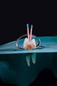
 Endodontic treatment, also known as root canal treatment, consists of removing the infected or inflamed nerve, sterilizing the root canal and filling it with a filling material to prevent reinfection and restore the tooth’s functionality.
Pulpal infections and inflammation can occur for many reasons. Untreated deep dental decay, dental trauma, dental fractures or cracks can damage the nerve of the tooth and lead to various pathologies. These conditions are quite common and can affect people of all ages. Common symptoms include spontaneous pain, sensitivity to hot or cold and during chewing, and in more complicated cases abscesses in the affected area.
Endodontic treatment involves several steps to treat and save the affected tooth. First of all, the dentist will identify the problem and assess the degree of damage to the nerve as well as the bone around the root. Then local anesthesia will be used to ensure comfort during treatment. Next, the root canal will be accessed, the affected pulp tissue will be removed and the root canal will be cleaned and disinfected with special solutions. After cleaning, the root canal will be filled with a filling material to prevent reinfection and to ensure complete sealing of the canal. Finally, depending on the degree of damage to the dental tissues, a dental crown may be applied to provide protection and restore functionality and aesthetics.
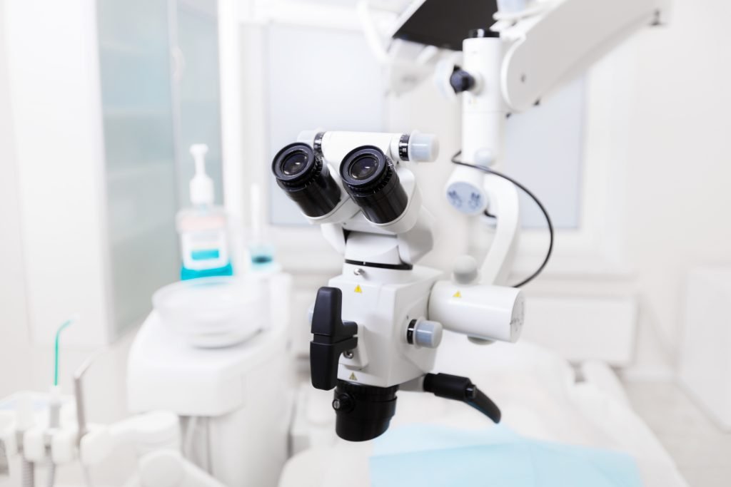
In the world of modern dentistry, the use of the microscope in endodontics has become an indispensable practice. This state-of-the-art instrument has revolutionized the approach to endodontic treatment, allowing dentists to achieve maximum precision and exceptional results.
What are the advantages of using a microscope in endodontics?
Visual magnification: The dental microscope provides a magnified view of the working area, allowing the dentist to observe the complex anatomy of the root canal and pulp tissue in detail. This magnified view is essential for identifying extra canals, root fractures and other critical anatomical details that may be overlooked with the naked eye.
Precision and accuracy: The endodontic microscope allows the dentist to perform endodontic procedures with extraordinary precision and accuracy. The magnified image and intense illumination help to pinpoint the exact location of root canals, eliminating the risk of losing or partially treating them. The microscope also facilitates complete removal of infected or damaged tissue, contributing to an optimal therapeutic outcome.
More accurate diagnosis: The microscope is not only useful during endodontic procedures, but also in the diagnostic phase. The magnified images provided by this device allow the dentist to better observe minor lesions or abnormalities of the tooth and root canal that may be difficult to detect with the naked eye. This precision in diagnosis ensures proper treatment planning and reduces the risk of subsequent failures or complications.